Fighting Cancer With Advanced Treatment & Compassion
Fighting Cancer With Advanced Treatment & Compassion
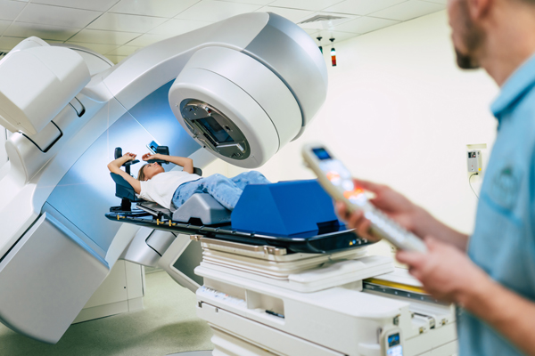
Advanced technology plays a pivotal role in transforming cancer care by enabling early detection, personalized treatment approaches, minimally invasive procedures, precise radiation therapy delivery, efficient data management & analysis, and remote patient monitoring. These advancements not only enhance clinical outcomes but also empower healthcare professionals with valuable tools to improve overall patient experiences throughout their cancer journey.
Precision in cancer care largely dependent on the type of technology that is used for diagnosing as well as treating cancer. At American Oncology Institute, one of the best cancer treatment hospitals in India, we are ahead of time in terms of both medical expertise and technology. All our centres are equipped with the most advanced technology and best-in-class machines. With the latest and advanced technology, we can precisely target cancer cells while minimizing damage to nearby healthy tissues—a vital aspect in reducing side effects during treatment.
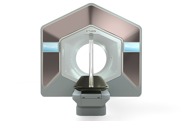
Ethos therapy integrated with IDENTIFY, a novel technology and technique provides the ability to alter the treatment plan by capturing the movement in internal and external anatomy during each treatment. This improves the pre-treatment plan and adapts to real-time changes during the treatment by gaining insights through the advanced power of AI-enhanced image segmentation and surface motion sensors. The Ethos Machine is a much safer and more reliable way to treat cancer patients than the conventional radiotherapy machines. It is designed to be used in combination with existing treatments, such as chemotherapy, targeted therapy, or immunotherapy. This allows for a more comprehensive approach to treating cancer, giving patients access to a range of treatment options. It is capable of targeting cancer cells in nearly any area of the body, including the brain, liver, and lungs. Adaptive therapy integrated with surface guided motion management helps to adapt the treatment plan and target the tumour based on the anatomical changes. The Ethos Machine is a revolutionary new treatment, capable of delivering a precise dose of radiation to target and kill cancer cells, while leaving healthy cells intact. The ability of the machine to deliver on-couch adaptive treatment puts the patient at the centre of care.
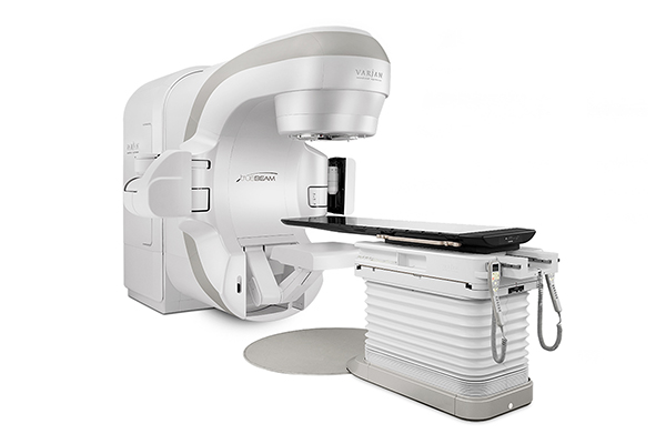
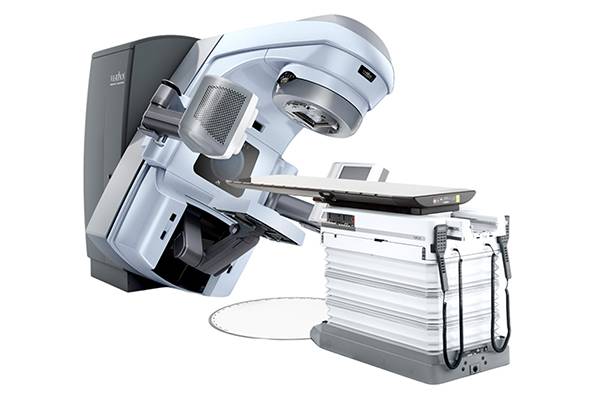
TrueBeam system makes it possible to deliver accurate image-guided treatments faster than was possible with earlier generations of Varian technology. In fact, most treatments take only a few minutes per day. Image-guidance tools can generate 3-D anatomical images 60% faster. These images can be generated using 25% less X-ray dose. This enables us to target tumors with accuracy that is measured in fractions of millimeters. Even tumors that move when a patient breathes (for example, those in the lung or breast) can be precisely targeted due to special tools that compensate for motion throughout a treatment. TrueBeam technology can be used for many forms of advanced treatments, including image-guided radiotherapy (IGRT), intensity-modulated radiotherapy (IMRT), RapidArc® radiotherapy and a new Gated RapidArc approach that makes it possible to compensate for tumor motion during a RapidArc treatment. With this level of flexibility, we can tailor treatments to a patient’s specific circumstances.
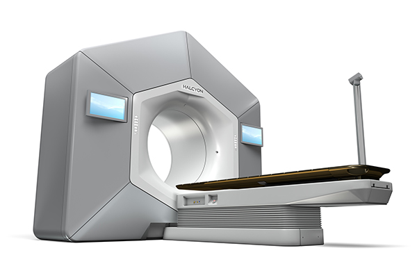
The Halcyon radiotherapy system is the most recent advancement from Varian, a leader in developing and delivering cancer care solutions. Its leading-edge technology precisely targets the tumor while providing a treatment environment that’s more comfortable than ever before. Halcyon was designed to provide treatment quickly and efficiently. Imaging can be completed in about 15 seconds, and brushless motors in the machine enable fast treatment delivery. With Halcyon, treatments are typically completed in just nine steps. That means your time on the machine is greatly reduced. In fact, most treatments are completed in about 10 minutes. That’s half of what’s expected with conventional machines. Halcyon delivers a precise, tightly focused radiation dose that conforms to the shape of your tumor while minimizing impact on surrounding healthy tissue. Halcyon can treat most disease sites, including the brain, head and neck, prostate, lungs, and breasts.
The Clinac iX is a state-of-the-art radiation therapy system developed by Varian Medical Systems. It is designed to deliver precise and effective radiation treatments for cancer patients. The Clinac iX utilizes advanced technologies such as Intensity-Modulated Radiation Therapy (IMRT) and Image-Guided Radiation Therapy (IGRT). This allows for highly accurate targeting of tumors while minimizing exposure to surrounding healthy tissues. The system offers a wide range of treatment techniques, including 3D conformal radiotherapy, IMRT, RapidArc®, stereotactic radiosurgery (SRS), and volumetric modulated arc therapy (VMAT). This versatility enables clinicians to tailor treatment plans based on individual patient needs. Integrated imaging systems provide real-time visualization throughout the treatment process, allowing for precise tumor localization and alignment before each session. This ensures that the radiation beam is accurately delivered to the intended target area. The system incorporates various features aimed at enhancing patient comfort during treatment sessions. These include advanced motion management technology that adjusts for respiratory or body movement, ensuring consistent delivery of radiation even with patient motion. Overall, the Clinac iX is a cutting-edge radiation therapy system that combines advanced treatment delivery techniques, precise imaging capabilities, and patient-centric features. It empowers healthcare professionals to p
High-Dose Rate (HDR) brachytherapy is a specialized form of radiation therapy used in the treatment of various types of cancer. It involves delivering a high dose of radiation directly to the tumor or the area surrounding it, using a temporary implant or applicator. With HDR brachytherapy, radioactive sources are placed inside or near the tumor for a short duration, allowing for precise and targeted delivery of radiation. This enables higher doses to be administered while minimizing exposure to healthy tissues. The treatment plan for HDR brachytherapy can be customized based on individual patient needs. The position and dwell times of the radioactive sources can be adjusted to deliver optimal radiation dose distribution within the tumor volume. By precisely controlling the placement and intensity of radiation sources, HDR brachytherapy minimizes damage to nearby healthy tissues compared to other forms of radiotherapy. The targeted nature of HDR brachytherapy helps minimize side effects compared to conventional radiation therapy. By limiting exposure to healthy tissues, patients may experience fewer complications and a faster recovery.
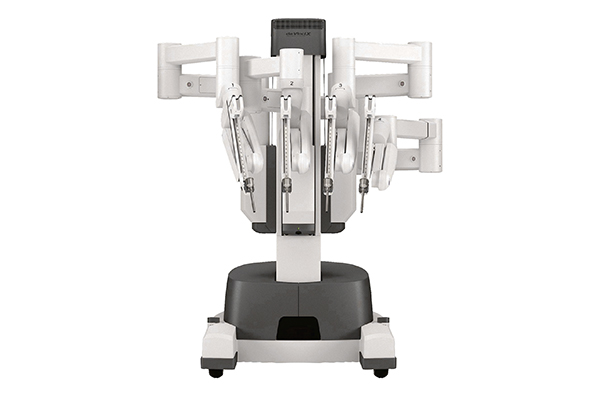
The da Vinci X System is an advanced robotic surgical system. It is designed to assist surgeons in performing minimally invasive procedures with enhanced precision and control. The da Vinci X System enables surgeons to perform complex surgeries using a combination of robotics, 3D visualization, and specialized instruments. It provides a high degree of dexterity, allowing for precise movements within the patient's body. With the da Vinci X System, surgeons can perform procedures through small incisions compared to traditional open surgery. This results in reduced trauma to surrounding tissues, less pain for patients, shorter hospital stays, faster recovery times, and smaller scars. The precision and control offered by the da Vinci X System can result in reduced blood loss, lower risk of complications, faster recovery times, and improved clinical outcomes for patients undergoing robotic-assisted surgeries. It's important to note that the da Vinci X System is not autonomous but rather enhances a surgeon's abilities with its advanced technology. Surgeons undergo specialized training to effectively utilize the system and ensure patient safety during procedures.
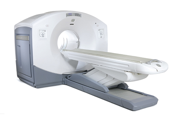
PET-CT, or Positron Emission Tomography-Computed Tomography, is a powerful imaging technique that combines two different scanning methods to provide detailed information about the structure and function of tissues within the body. The integration of PET and CT scans into one examination allows for precise localization by merging functional data from PET with anatomical details from CT imaging. This fusion enhances diagnostic accuracy by providing comprehensive information on both physiological changes (PET) along with their precise anatomical location (CT). PET-CT is particularly valuable in oncology as it can detect areas of abnormal metabolic activity associated with cancer cells throughout the body—helping identify primary tumors, metastases, or recurrence. It plays a crucial role in cancer staging by assessing disease spread accurately. PET-CT has revolutionized medical imaging by combining functional and anatomical data into one comprehensive examination. Its versatility across various medical specialties contributes significantly to improved patient management—from early detection and accurate staging of diseases to treatment planning and monitoring responses over time.
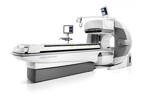
SPECT-CT, or Single Photon Emission Computed Tomography-Computed Tomography, is an advanced imaging technique that combines two different scanning methods to provide comprehensive information about the structure and function of tissues within the body. The integration of SPECT and CT scans into one examination allows for precise localization by merging functional data from SPECT with anatomical details from CT imaging. This fusion enhances diagnostic accuracy by providing comprehensive information on both physiological changes (SPECT) along with their precise anatomical location (CT). SPECT-CT plays a valuable role in oncology by helping identify areas of abnormal metabolic activity associated with cancer cells—allowing detection, characterization, staging, and monitoring treatment response. It aids in determining tumor size, extent, spread, as well as identifying potential sites for biopsy or surgery planning. SPECT-CT has revolutionized medical imaging by combining functional data from SPECT with precise anatomical information from CT scans into one comprehensive examination. This integration enhances diagnostic accuracy across various medical specialties—from oncology and cardiology to neurology and musculoskeletal imaging—enabling targeted treatment plans tailored to individual patient needs.
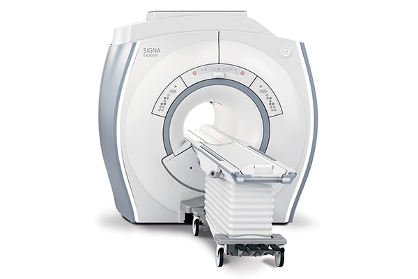
MRI, or Magnetic Resonance Imaging, is a non-invasive imaging technique that uses powerful magnets and radio waves to generate detailed images of the body's internal structures. The term "1.5 Tesla" refers to the strength of the magnetic field used in the MRI scanner. A 1.5 Tesla MRI scanner utilizes a moderate-strength magnetic field for image acquisition. While higher-field strength scanners exist (e.g., 3 Tesla), a 1.5 Tesla magnet is still widely used and offers excellent image quality for most diagnostic purposes. MRI provides highly detailed cross-sectional images of various tissues within the body, including organs, muscles, bones, blood vessels, and nerves. A 1.5 Tesla magnet allows for high-resolution imaging that can aid in accurate diagnosis by visualizing even small anatomical structures. MRI with a 1.5 Tesla magnet is suitable for examining various parts of the body, such as brain, spine, joints (e.g., knee, shoulder), abdomen/pelvis, chest, etc. It enables evaluation across different medical specialties—from neurology and orthopedics to oncology and cardiology. One major advantage of MRI is its ability to differentiate between different types of soft tissues based on their inherent properties (e.g., water content). This makes it particularly useful in identifying abnormalities or distinguishing between normal/abnormal tissues—for example, detecting tumors or evaluating joint injuries.
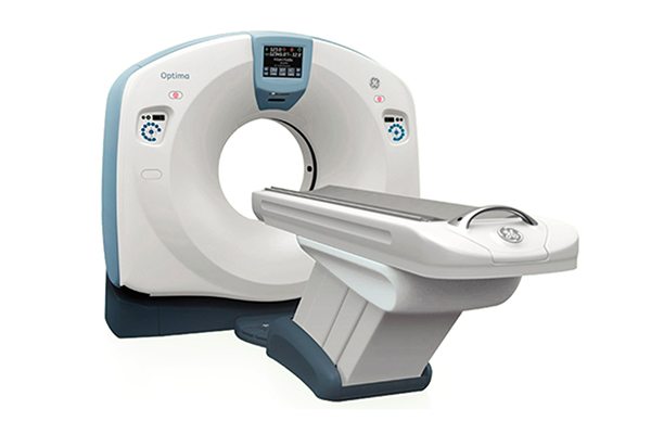
CT, or Computed Tomography, is a medical imaging technique that uses X-rays and advanced computer processing to create detailed cross-sectional images of the body. CT provides highly detailed images of internal structures such as bones, organs, blood vessels, and soft tissues. It offers excellent visualization of anatomical details in multiple planes (axial, sagittal, coronal), allowing for accurate diagnosis. CT scans can be completed quickly—typically within minutes—which is especially beneficial for patients who require urgent medical evaluation or those who may have difficulty remaining still during longer procedures. This makes it a valuable diagnostic tool across multiple medical specialties—from emergency medicine and oncology to orthopaedics and neurology. CT has revolutionized medical imaging with its ability to generate detailed cross-sectional images quickly and accurately. Its versatility across different body regions and applications makes it a go-to modality for diagnosis, treatment planning, and monitoring response to therapy across numerous medical specialties.
Life does not stop when cancer strikes.
We are with you in this fight to win over cancer. We are here to give you the strength to recover through a comprehensive cancer care program.